
Figure 2.
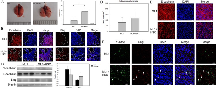
HSCs enhance ML1 metastasis to lung in NOD/SCID mice with EMT induction. The hepatoma cell line ML1 was subcutaneously injected (1x10⁶) into NOD/SCID mice with or without same amount (1x10⁶) of HSC-T6 cells. The mice were sacrificed at 6 weeks post injection. A. A representative lung tissue picture and the number of lung metastatic nodules was significantly higher in the mice of ML1 with HSC; B. the expression of E-cadherin (red) was down-expressed while the expression Slug (red) was increased in the mice with of ML1 with HSC when compared to the mice with of ML1 only; C. The immunoblotting of E-cadherin, N-cadherin, and Slug protein in lung tissue showed similar result; D. The subcutaneous tumor size was measured at sacrifice time point; E and F. The immunohistochemistry staining of E-cadherin (red), α-SMA (green), and Slug (red) in subcutaneous tumor sites. The cell nuclei were stained with DAPI (blue). Each group contains 6-8 mice. P < 0.05 were considered significant.