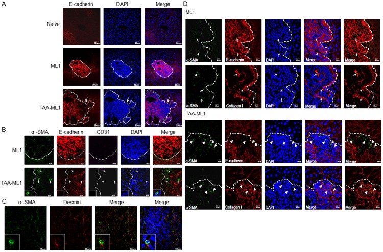
Figure 6.
Hepatic stellate cells were localized around E-cadherin down-expressed tumor region in the liver fibrosis combined with hepatoma model. A. E-cadherin (red) is highly expressed in DAPI (blue) condensed tumor region (dot line) harvested from hepatoma bearing (ML1) BALB/c mice. In the fibrosis combined with hepatoma (TAA-ML1) mice liver, E-cadherin is down-expressed in front parts of the tumor region as indicated by an arrow. (200x); B. The hepatic stellate cells in tumor were stained with α-smooth muscle actin (α-SMA, green). Anti-E-cadherin (red), anti-CD31 (purple, triangle), and DAPI (blue) were also stained. The tumor regions with high α-SMA expressed cells were E-cadherin down-expressed; C. These cells were desmin+ but CD31-. (600x) The images were representative to 5-7 mice in each group; D. The mice liver with hepatoma (ML1) or hepatoma combined with fibrosis (TAA-ML1) were collected and sliced into 5 μm sections. Immunofluorescence staining of α-smooth muscle actin (α-SMA, green), E-cadherin (red), collagen type I (red), and DAPI (blue) in liver tissue section. The dotted line shows the tumor area. Arrowhead indicates the α-SMA highly expressed cells, and arrow indicates the α-SMA+ cells with lower expression.

