Figure 6.

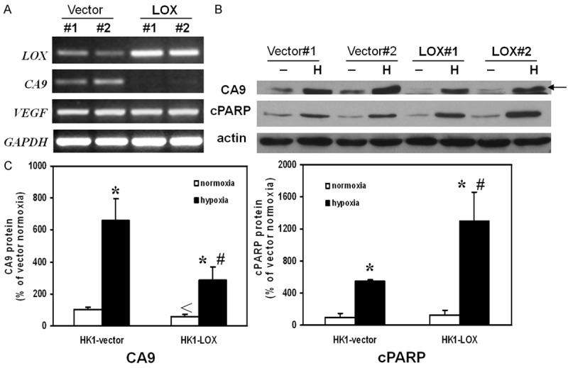
RT-PCR and Western Immunoblotting analysis. A. Expression of LOX in HK1 cells transfected with LOX plasmid was confirmed. Expression of CA9 was down-regulated in LOX plasmid transfected HK1 cells while the expression of VEGF was not altered. B. Overexpression of LOX altered protein levels of CA9 and cPARP under hypoxia treatment. Representative photos were taken from three independent experiments. C. Quantitative analysis of CA9 protein and cPARP protein by densitometry. The value of band intensity from HK1-vector under normoxia was expressed as 100%. Results were depicted as means ± SEM of three independent experiments. The * indicates statistical significant when compared to same cells under same treatment condition. The # indicated statistical significant when compared to vector control during hypoxia conditions. The < indicates statistical significant when compared to vector control during normoxia conditions.
