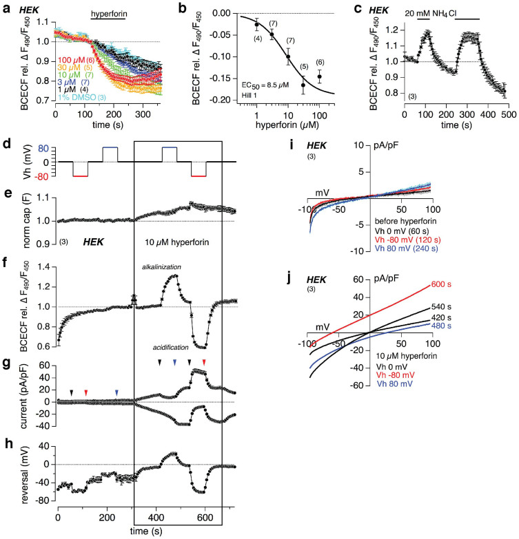
Figure 4. Hyperforin induces changes of the intracellular pH in HEK cells.
(a, c) Relative changes of the fluorescence ratio (F490/F450) of the pH-sensitive dye BCECF-AM in HEK cells. As indicated by the bars different concentrations of hyperforin or 1% DMSO (a), or 20 mM NH4Cl (c) were applied. External NH4Cl induces intracellular alkalization and thus increase of F490/F450 (c). The sigmoidal fit of the relative decrease of the BCECF ratio at different hyperforin concentrations, calculated at 200 s in respect to the control application of DMSO, reveal a half-maximal concentration (EC50) of 8.5 μM (b). Normalized capacitance (e), intracellular pH changes (F490/F450; f), inward and outward currents at −80 mV and +80 mV, respectively, normalized to the cell size (g), and reversal potential (h) before and during 10 μM hyperforin, measured in HEK cells with the free acid of BCECF in the patch pipette. (d) depicts the changes of the holding potential (Vh), providing the driving force for the currents. IVs, extracted at the indicated time points (see arrowheads in g) before and during 10 μM hyperforin, are displayed in (i) and (j), respectively. Data represent means ± S.E.M. of the indicated number of experiments (cells).

