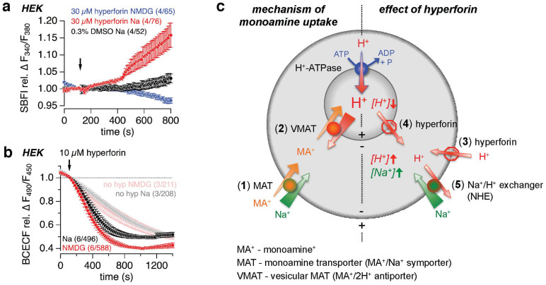
Figure 7. Model of hyperforin action on monoamine uptake.
(a, b) Relative changes of the SBFI fluorescence ratio (F340/F380), representing intracellular Na+ changes (a), and the BCECF fluorescence ratio (F490/F450), representing intracellular pH changes (b), in HEK cells during the application (see arrowhead) of 30 μM (a) and 10 μM (b) hyperforin in the presence and absence of Na+ (replaced by NMDG+). In (a) 0.3% DMSO was applied as control. In (b) control experiments without application of hyperforin are shown in faint colors. Data represent means ± S.E.M. of the indicated number (n) of experiments including x cells (n/x). (c) Mechanism of monoamine uptake (left side of the model): Cellular (synaptic) uptake of monoamines (MA+) is arranged by the plasma membrane monoamine-sodium symporter (MAT; (1)). The negative membrane potential and low intracellular Na+ concentration provide the driving force for cellular Na+ and MA+ uptake. Vesicular monoamine uptake is arranged by the vesicular monoamine-proton antiporter (VMAT; (2)), moving MA+ into the vesicle in exchange for two protons. The huge H+ gradient and the vesicular membrane potential, both established by the vesicular proton pump (H+-ATPase) drives H+ out of the vesicle and thereby promotes vesicular MA+ uptake. Effects of hyperforin on monoamine uptake (right side of the model): The protonophore action of hyperforin moves H+ into the cell (synapse) due to the negative membrane potential (3). In addition, hyperforin moves H+ out of the vesicles, driven by the huge H+ gradient and positive vesicular membrane potential (4). The dissipation of the vesicular H+ gradient ruins the driving force for the vesicular VMAT-dependent MA+ uptake (2). The cytosolic acidification, mediated by the hyperforin-dependent H+ influx and H+ release from vesicles, increases the activity of the plasma membrane sodium-proton exchanger (NHE; (5)), which is driven by the Na+ gradient and the negative membrane potential, and moves one H+ out of the cell in exchange of one Na+ into the cell. This results in an increase of cytosolic Na+ concentration, reducing the driving force for the MAT-dependent cellular MA+ uptake (1).

