
Figure 1. Structural changes and sequence alignment of the G5 loop.
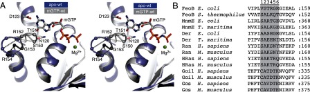
(A) Stereo view of the G5 loop in EcNFeoB. Nucleotide free (PDB code 3HYR) and nucleotide bound (3HYT) structures are shown in purple and grey, respectively. The residues in the G5 loop and selected residues involved in nucleotide base coordination are labelled and shown as spheres and ball-and-stick, respectively. The conformational shift of individual Cα atoms in the G5 loop is illustrated with dotted lines. GMPPNP is shown as ball-and-stick, with the mant group removed for clarity. (B) Sequence alignment of the residues in and around the G5 sequence motif. Residues numbered and shaded in grey are, in this study, designated as G5 loop residues.