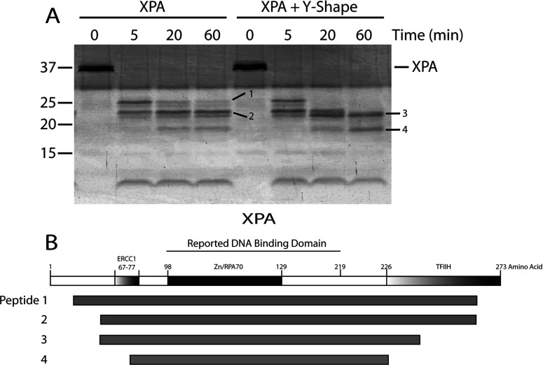
Figure 4. Partial proteolysis of XPA versus XPA–DNA junction complex by chymotrypsin.
(A) Four microgram of XPA was digested with chymotrypsin (1:80 molar ratio of chymotrypsin:XPA) at room temperature for the times indicated. The reactions were terminated and the cleavage products resolved by SDS–PAGE (15% gel) then visualized using SYPRO Ruby stain. Untreated XPA was loaded as a control (0 time). The molecular mass markers are indicated on the left. Individual fragments of interest are designated by dashes with numbers. (B) Schematic map of XPA with its functional domains highlighted followed by a schematic of the four proteolytic peptides indicated in (A) that were generated upon XPA treatment with chymotrypsin.

