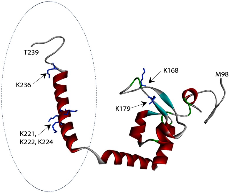
Figure 5. A structural model of redefined DBD of XPA.
Structure of the previously reported DBD (aa98-219) of XPA is shown with the predicted extended-structure generated by RaptorX (aa217–239, indicated by dashed circle). Structural mode of the redefined DBD of XPA was generated by Discovery Studio tethering of the two structures together by overlapping three amino acids shared between the two structures. Lysine residues in the structure are presented in stick representation. K168 and K179 are found in the previously reported DBD of XPA, while K221, K222, K224 and K236 are found within the extended structure. The two structures resemble two arms of a clamp. Biotin-modified lysine residues protected from modification in the presence of ds–ssDNA junctions are shown in blue.

