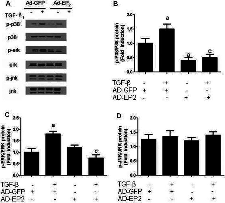
Figure 11. The expression of p-p38MAPK, p-ERk and p-JNK in AP-EP2-infected MCs.
Primary MCs were infected with AP-EP2 for 24 h and then incubated with 10 ng/ml TGFβ1 for 24 h. The expression of p-p38MAPK, p-ERK and p-JNK in AD-EP2+TGFβ1 group were significantly decreased than that of AD-GFP+TGFβ1 group. (A) Western Blot. (B–D) Quantification of p-p38MAPK, p-ERK and p-JNK expression is achieved using densitometric values normalized to β-actin levels (aP<0.05 versus AD-GFP group; cP<0.05 versus AD-GFP+TGFβ1 group).

