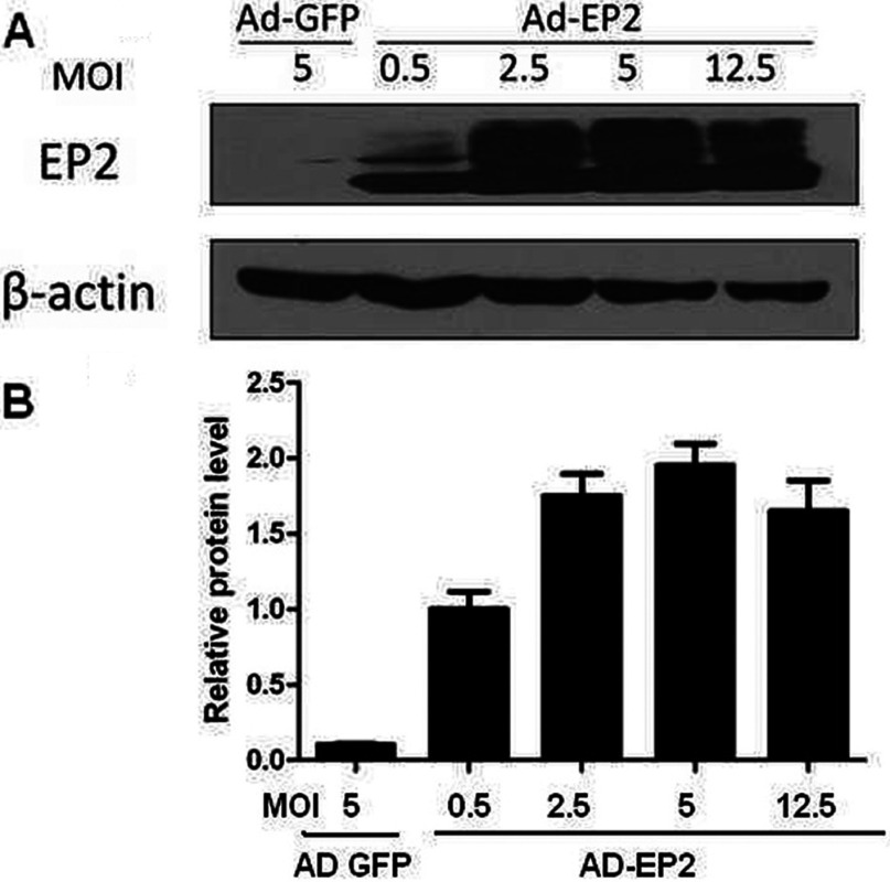
Figure 3. Expression of EP2 in AD-EP2 infected mouse MCs.
Primary mouse MCs were infected with AD-GFP or different concentration of AD-EP2 (MOI=0.5, 2.5, 5, 12.5) for 24 h and then EP2 protein level was detected by Western blot analysis, β-actin was used as control. (A) Expression of EP2 in AD-EP2 infected MCs was detected by Western blot. (B) Quantification of EP2 expression is achieved using densitometric values normalized to β-actin levels.

