
Fig. 1.
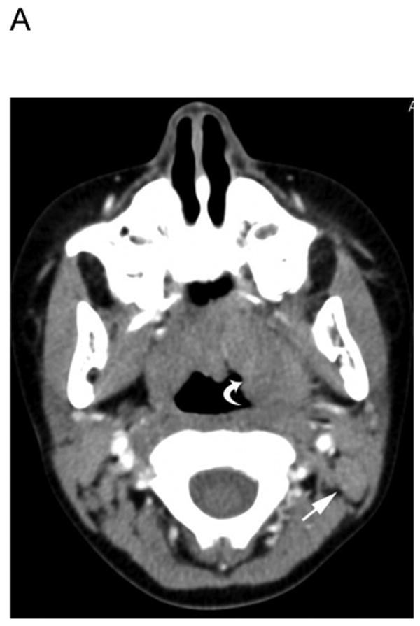
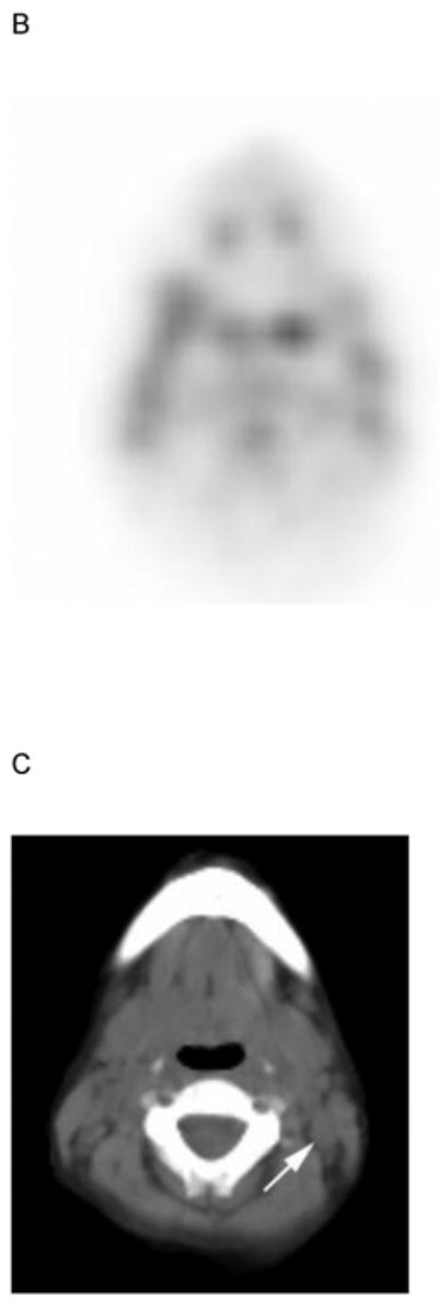
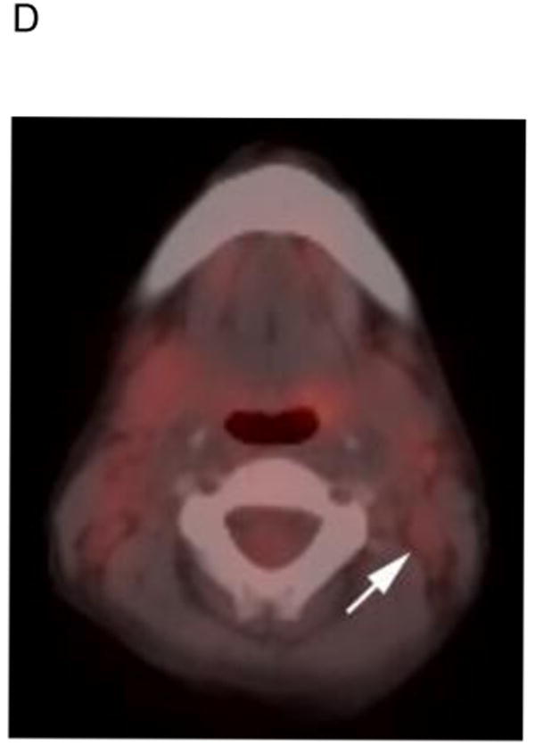
5 yo girl with left-sided nasopharyngeal rhabdomyosarcoma (not otherwise specified). A) Diagnostic computed tomography (CT) shows a 1.4 cm left posterior cervical lymph node (arrow) that was concerning for metastasic disease. Note primary tumor in left nasopharyngeal tonsil (curved arrow). B) Axial positron emission tomography (PET) image, C) co-registered non-diagnostic CT and D) fused PET-CT images show minimal uptake within the suspicious node (arrow) that was symmetric with the opposite side and interpreted as benign. The patient had no nodal recurrence or other evidence of nodal disease.