
Figure 3.
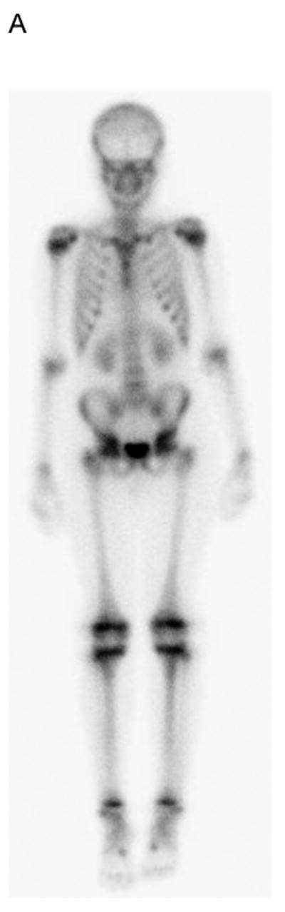
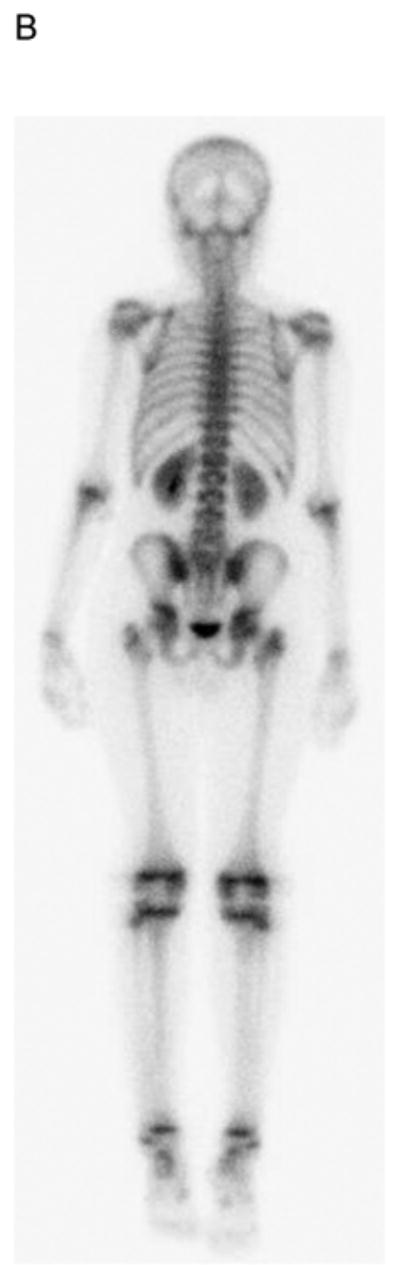
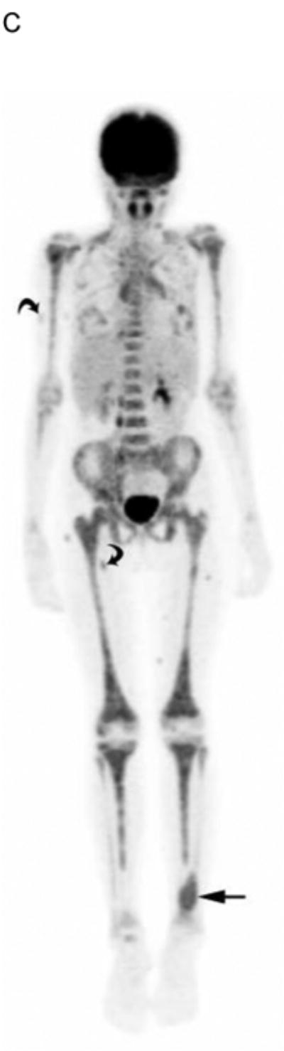
19 yo girl with embryonal RMS, primary site left lower leg. A) Anterior and B) posterior 99mTc bone scan images show no evidence of bone metastasis. C) Maximum intensity projection (MIP) PET-CT image showing diffusely abnormal and mildly asymmetric marrow FDG uptake throughout the upper and lower extremities, pelvis and spine, due to marrow disease proven by biopsy. The primary left lower leg tumor (straight arrow) and soft-tissue metastases (several indicated with curved arrows) are also evident. The soft tissue metastases were not detected by CI or physical examination.