
Figure 3.
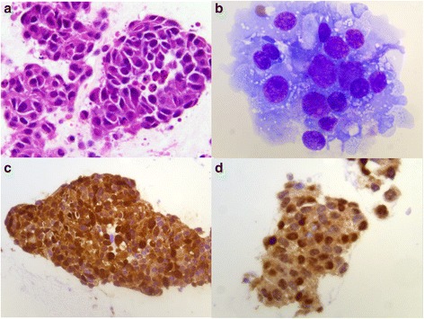
Histopathology. Photomicrograph shows cohesive groups of malignant epithelioid cells displaying pleomorphic round eccentric nuclei with prominent nucleoli and abundant cytoplasm. a) Hematoxylin and eosin, 63× b) Diff-Quik, 100× . Immunohistochemistry discloses positive staining for c) S-100 stain, 40× and d) microphthalmia stain, 63×.