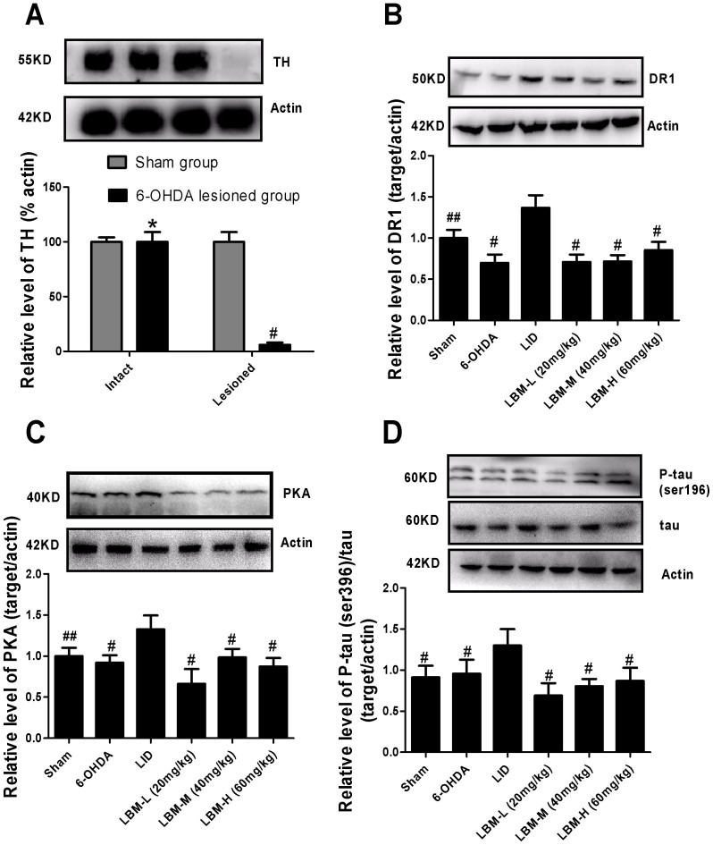
Figure 5. LBM prevented increased levels of D1R, PKA, p-tau and total tau after pulsatile L-dopa treatment.
Protein levels were evaluated by western blotting of proteins extracted from the ipsilaterally striatum of the rat brains. They were assessed in extracts from sham or 6-OHDA-lesioned rats treated with vehicle, pulsatile L-dopa (20 mg/kg, bid) or LBM-L (20 mg/kg, sc), LBM-M (40 mg/kg, sc) and LBM-H (60 mg/kg, sc). (A) Extent of the dopaminergic denervation induced by 6-OHDA lesions and sham. Tyrosine hydroxylase (TH) levels expressed relative to actin levels. # p < 0.01 and * p > 0.05 vs sham-intact hemisphere (n = 4); (B) DR1 levels expressed relative to actin levels; (C) PKA levels expressed relative to actin levels; (D) phosphorylated tau levels at ser396 expressed relative to tatal tau levels. The data represent the mean of relative optical density (expressed as a percentage of respective control values and normalized using sham group) ± SD; # p < 0.01 and ## p < 0.05 vs LID group (n = 4).

