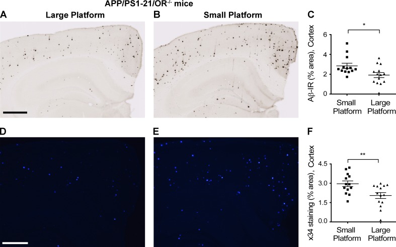
Figure 7.
Increased Aβ deposition with chronic sleep deprivation in APP/PS1-21/OR−/− mice. (A–F) The amount of Aβ plaques stained with HJ3.4B (A–C) and fibrillar Aβ stained with X-34 (D–F) were compared between APP/PS1-21/OR−/− mice exposed to a large platform and a small platform. Sleep deprivation experiments were performed using a small and large platform in a cage with water on the bottom, where a mouse cannot sleep on the small platform because of its size, whereas they can maintain a normal sleep–wake cycle on the larger platform. Mice exposed to small platforms (n = 3) and to large platforms (n = 3) were analyzed together within a set of experiments. The results are the sum of five repeats in different mice. Each mouse was investigated independently. Data are presented as mean ± SEM (n = 13–14 in each group, two-tailed Student’s t test). *, P < 0.05; and **, P < 0.01. Bars: (A) 500 µm; (D) 100 µm.

