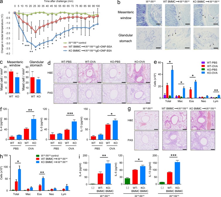
Figure 5.
Systemic anaphylaxis and the OVA-induced allergic response in mast cell–reconstituted Wsh/Wsh mice. (a) Mast cell–deficient mice were given no BMMCs (Wsh/Wsh control; n = 2) or were reconstituted with BMMCs from Tespa1 WT mice (WT BMMC→Wsh/Wsh; n = 6) or KO mice (KO BMMC→Wsh/Wsh; n = 6). 16 wk later, mice were sensitized with 10 µg anti-DNP IgE, followed by challenge with 100 µg DNP-BSA. Rectal temperature was monitored every 5 min. (b) Tissue mast cells in the mesenteric window and glandular stomach from the indicated chimeric mice were assessed toluidine blue staining. Arrowheads indicate individual mast cells. Bars, 100 µm. (c) Absolute numbers of mast cells per mm2 (mean and SD). WT and KO mice (n = 6 mice per group; d–f) or chimeric mice (n = 5 per group; g–i) were sensitized with OVA. Mice were challenged with OVA or PBS in the airway inflammation model and analyzed 24 h after the final challenge. (d and g) Sections of lung tissues were stained with hematoxylin and eosin (H&E). Bars, 100 µm. (e and h) Numbers of total cells (total), eosinophils (Eos), neutrophils (Neu), lymphocytes (lym), and macrophages (Mac) in BALF. (f and i) Amounts of IL-4, IL-5, and IL-13 in BALF as measured by ELISA. Data are representative of two independent experiments. *, P < 0.05; **, P < 0.01; ***, P < 0.001 (Student’s t test).

