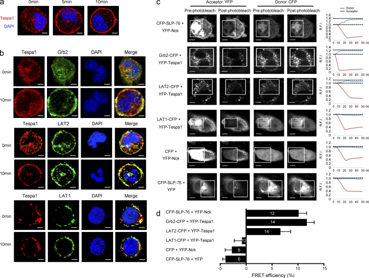
Figure 7.
Involvement of Tespa1 in FcεRI-mediated LAT2 signalosome assembly and preferential binding of Tespa1 with LAT2 versus LAT1. (a) BMMCs were sensitized with anti-DNP IgE and labeled with anti-Tespa1 (red) and DAPI (blue) before (0 min) and after (5 and 10 min) of stimulation. Bars, 2 µm. (b) BMMCs stimulated as in a were labeled with anti-Tespa1 (red), anti-Grb2 (green, top group), LAT2 (green, middle group), or anti-LAT1 (green, lower group) and DAPI (blue) before (0 min) and after (10 min) stimulation. Bars, 2 µm. (c) Live COS-7 cells transfected with the indicated pairs of CFP- and YFP-bearing constructs and analyzed by acceptor photobleaching FRET microscopy. White boxes indicate the bleached region. Group (right), donor, and acceptor relative fluorescence intensities were monitored in the bleached regions and plotted over time. Bars, 15 µm. (d) Average FRET efficiency obtained from acceptor photobleaching. White numbers in the bar graph indicate the numbers of cells used for FRET measurement. Detectable FRET between CFP-SLP-76 and YFP-Nck was taken as positive control; little or no detectable FRET between YFP-Nck and free CFP or between CFP-SLP-76 and free YFP were taken as negative controls. Data are representative of at least three experiments.

