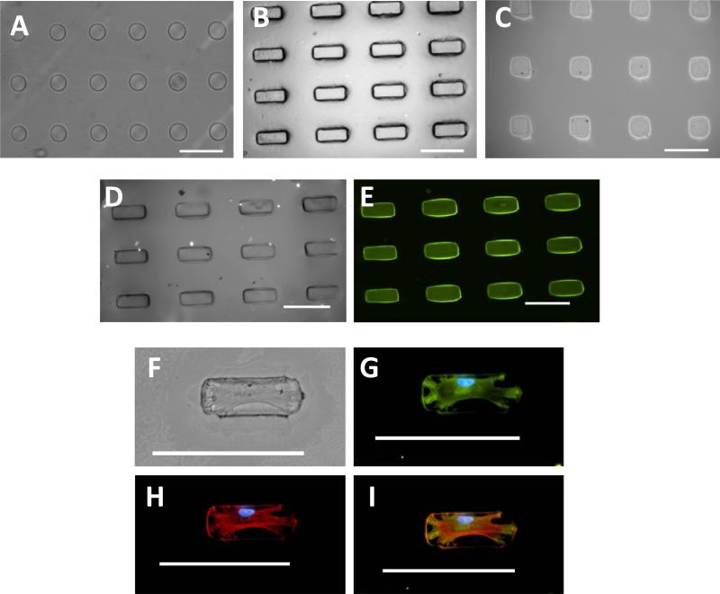
Figure 4.
Hydrogel islands were fabricated with protein exclusively attached to islands facilitating cell adhesion. Presented are microscopy images with micropatterned shapes showing (A) 5,000 μm2 circles (B) 5,000 μm2 rectangles and (C) 5,000 μm2 squares. Fluorescent bovine serum albumin was used as a model protein to determine protein attachment to micropatterned areas with (D) brightfield microscopy image of 5,000 μm2 rectangles and (E) bovine serum albumin exclusively attached to hydrogel rectangles. MSC attachment shown with (F) brightfield microscopy and immunofluorescence stained for (G) vinculin to reveal focal adhesions, (H) F-actin, and (I) merged image. Scale bars are 100 μm.

