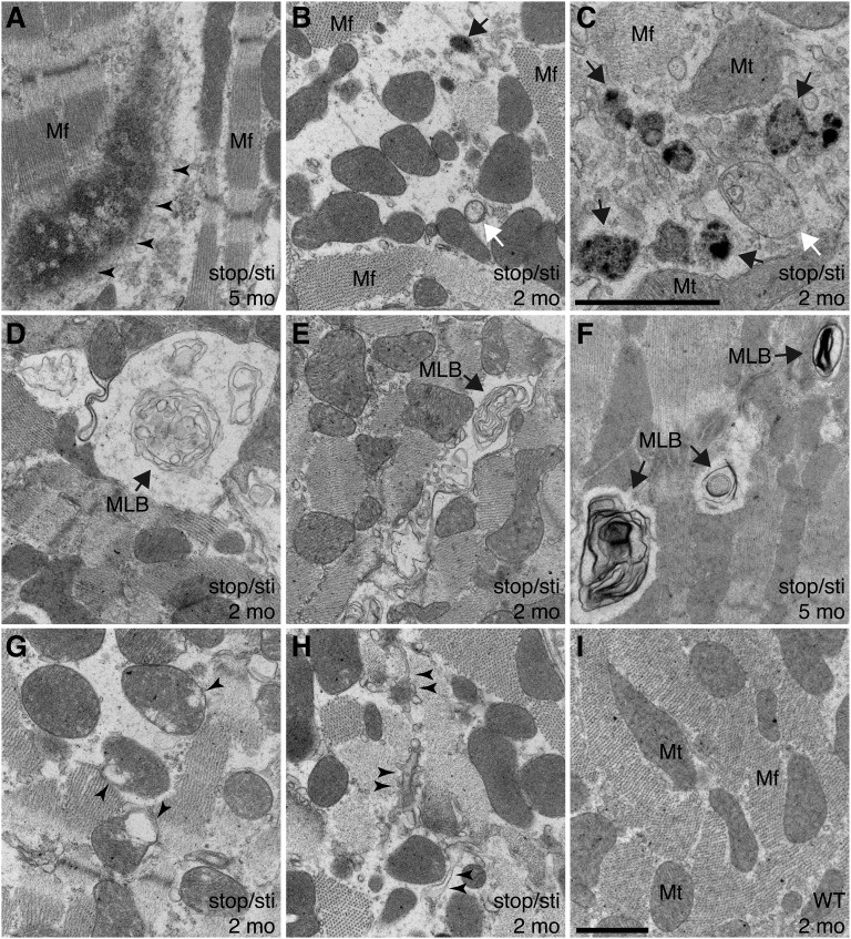
Fig. 4.
Ultrastructural changes in Aarsstop/sti cardiomyocytes. (A–I) Representative transmission electron microscope images of hearts from 2-mo- (B–E, G, and H) and 5-mo-old (A and F) Aarsstop/sti mice and 2-mo-old WT (I) mice. (A) Formation of large protein aggregates (arrowheads) that disrupt myofibril (Mf) structures. (B and C) Accumulation of autophagic vacuoles (white arrow, early autophagosome; black arrow, autolysosome) accompanied by loss of Mf structures. (D–F) Multilamellar bodies (MLBs, arrows), products of active autophagy and a hallmark of insufficient lysosomal activity, are also present. (G) Defective mitochondria (Mt) with disrupted cristae (arrowheads). (H) Disrupted sarcoplasmic reticulum (arrowheads) in Aarsstop/sti cardiomyocytes. [Scale bars, (A, B, and D–I) 1 μm and (C) 1 μm.]

