Fig. 6.
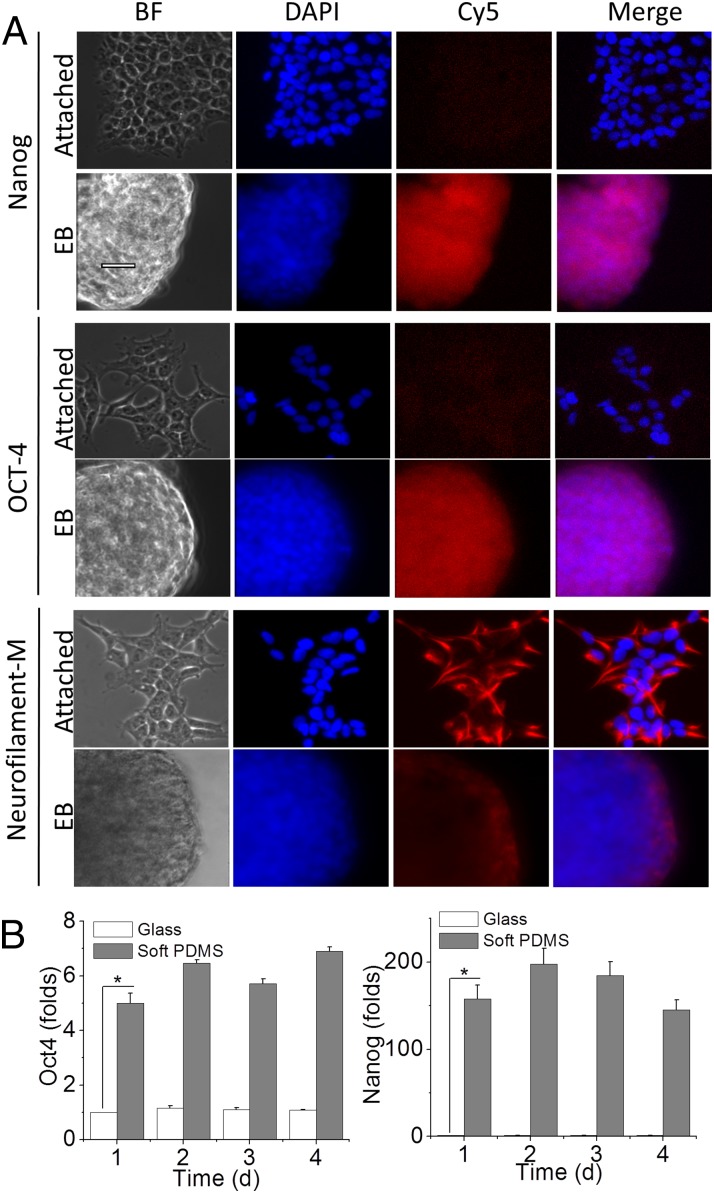
Stem cell markers in HEK-derived EBs. (A) HEK cells cultured on a coverslip (attached) and PDMS (EB, embryonic body) were immunostained with anti-Nanog, OCT4, and Neurofilament-M antibodies. Then, the cells were treated with secondary antibodies conjugated with Cy5. The nuclei were stained with DAPI. BF, bright field. (Scale bar, 50 μm.) (B) Real-time RT-PCR of HEK cultured on coverslip glass and soft PDMS for 1, 2, 3, and 4 d. The histogram shows the average fold of increase of OCT4 and Nanog from three sets of independently prepared samples at each time point (*P < 0.05 by Student t test).

