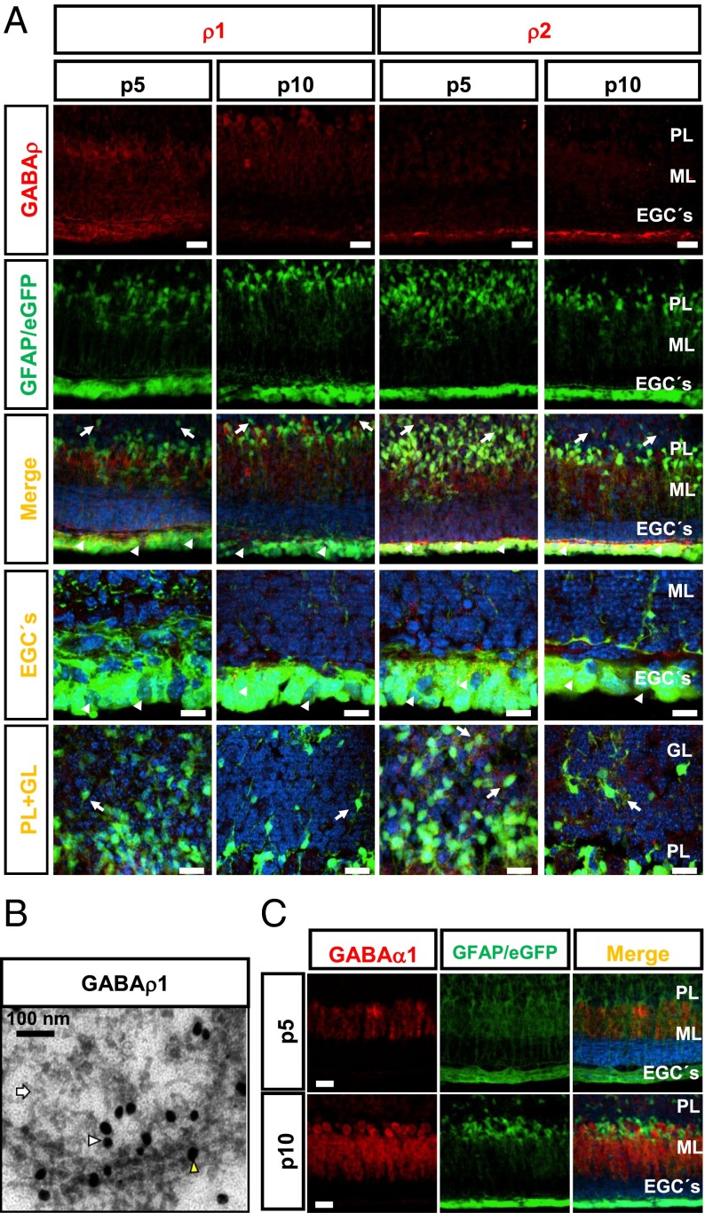
Fig. 1.
Expression of GABA-A receptor subunits in cerebellar GFAP+ cells. (A) GABAρ1 or GABAρ2 (first row) at P5 and P10 in GFAP-GFP transgenic mice (second row). The merged image shows that GFAP+ cells (green) of the subventricular zone (arrowheads) and GL (arrows) express GABAρ1 and GABAρ2 (red). The last two columns show maximizations of EGCs and GFAP+ cells of the GL expressing GABAρ. (Scale bars: 50 μm.) (B) Ultrastructural location of GABAρ1 in glial cells of the GL of cerebellum. The yellow arrowhead points to a gold particle labeling GABAρ1 in the plasma membrane; the white arrowhead, to a GABAρ1 label in submembranous structures. Astrocytes were identified by the presence of filamentous structures (white arrows). (Scale bar: 100 nm.) (C) Expression of GABAα1 (red) at P5, but not at P10, is located in Bergmann glial processes (green). n = 3. ML, molecular layer; PL, Purkinje layer. (Scale bars: 50 μm; 20 μm for maximizations.)

