Fig. 4.
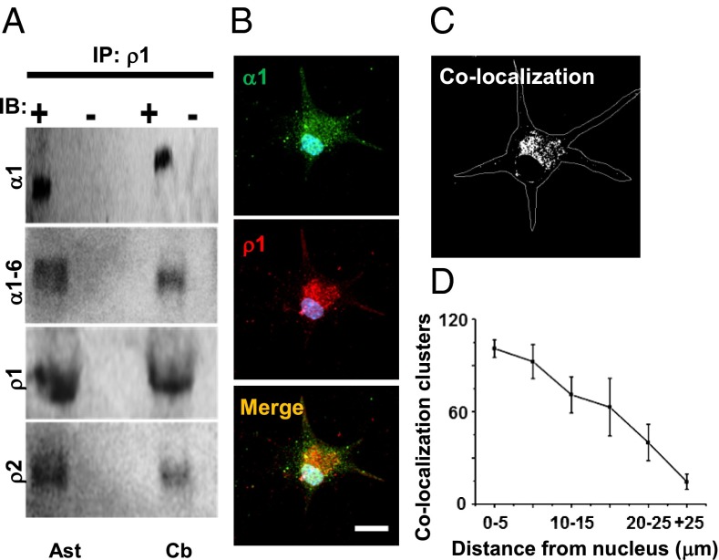
Protein–protein interaction between GABAρ1 and GABAα subunits. (A) Coimmunoprecipitation using antibody against GABAρ1 showing interactions among GABAα1–6-GABAρ1; GABAα1-GABAρ1, and GABAρ1-GABAρ2 subunits in cerebellar astrocytes (Ast) in culture and in adult cerebellum (Cb). Membrane proteins without anti-GABAρ1 were negative. (B) Immunolocalization of GABA-Aα1 (green) and GABAρ1 (red). Merged yellow clusters indicate colocalization of GABAα1 and GABAρ1. (C) Fluorescence from clusters colabeled for GABA-Aα1 and GABAρ1 converged mainly at the plasma membrane around the soma, and to a lesser extent in the distal processes of GFAP+ cells in culture (450 clusters from five cells). (D) Quantification of colabeled clusters.

