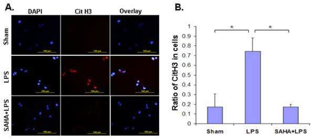
Figure 1. SAHA suppresses LPS-induced Cit H3 production.
(A) A representative CitH3 staining. (B) Ratio of CitH3 positive cells to all cells. Cell culture and immunostaning are described in Materials and Methods. The red color denotes decondensed chromatin stained with the Cit H3 antibody. 4'-6-Diamidino-2-phenylindole (DAPI) was used for nuclei staining (blue color). Statistical analysis shows that SAHA significantly suppressed the LPS-induced Cit H3 production (n=3; p<0.05).

