
Figure 4.
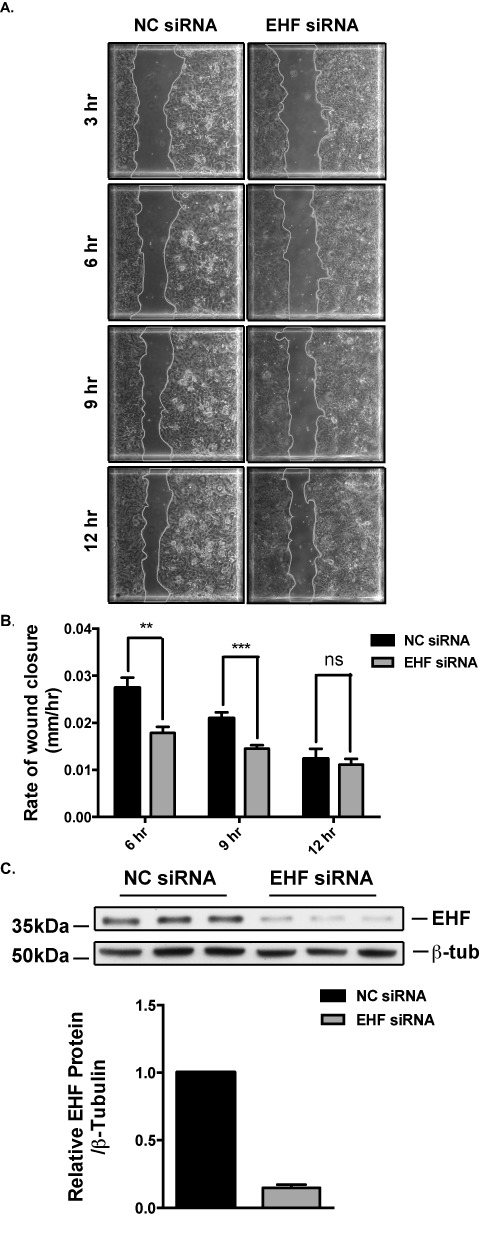
Effect of EHF depletion on wound repair in Calu-3 cells. (A) Images of negative control (NC) and EHF siRNA treated cells at 3, 6, 9 and 12 h after wounding. The outline of the wound is traced with a gray line. (B) Depletion of EHF significantly reduced the rate of wound closure at 6 and 9 h after wounding (n = 3). This difference was no longer evident 12 h after wounding. **P<0.01, ***P<0.001. (C) siRNA knockdown of EHF was measured by western blot in lysates taken 27 h after wounding and the signal was quantified using densitometry. EHF depletion was maintained throughout the experiment.