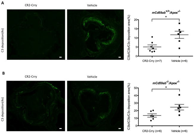
Figure 3. C3b/iC3b/C3c deposition was reduced in atherosclerotic lesions of CR2-Crry-treated mice.
A and B, C3b/iC3b/C3c deposition in aortic root of mCd59a+/+/Apoe−/− (A) and mCd59ab−/−/Apoe−/− mice (B) (Left, CR2-Crry; Right, PBS). The mean of the quantitative results of three sections obtained from each mouse was used to perform the statistical analysis. Scatter plot graph shows levels of C3b/iC3b/C3c deposition (percentage of positive area vs. lesion area) detected in atherosclerotic lesions. Scale bar: 100μm *P<0.05.

