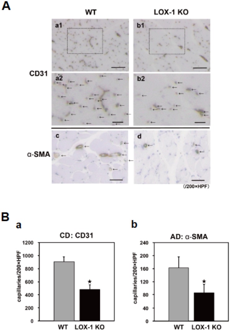
Figure 2. Determination of capillary density (CD) and arteriole density (AD).
A: Representative photographs to show CD and AD evaluated by histological examination of 20 randomly selected fields of tissue sections. CD in the ischemic tissues was immunostained with CD31 (an endothelial cell marker; brown, a1,2 and b1,2 and arrows, a2 and b2) and AD was anti-α-smooth muscle actin (α–SMA: an arteriole marker; brown and arrows, c and d) in the ischemic tissues of the gastrocnemius muscles on postoperative day 14 in high-power microscopic fields (x200). The number of vessels was reduced in LOX-1 KO mice (b1,2) compared with that in WT mice (a1,2) in CD31 staining (arrows in a2 and b2: the area of a1 and b1 surrounded by dotted line) and the number of arterioles stained with α–SMA (arrows in c and d) was also reduced in LOX-1 KO mice (d) compared with that in WT mice (c), Original bars; a1, b1: 300 µm a2, b2: 100 µm, c, d: 300 µm. B: CD and AD (mean±SD) were quantitatively assessed by histological examination of 20 randomly selected fields of tissue sections stained with CD31 staining (a) and α–SMA staining (b). The calculated capillary and arteriole density (capillaries per x200 HPF, arterioles per x200 HPF) were significantly lower in LOX-1 KO mice than in WT mice. ★, P<0.01 vs. WT, n = 20.

