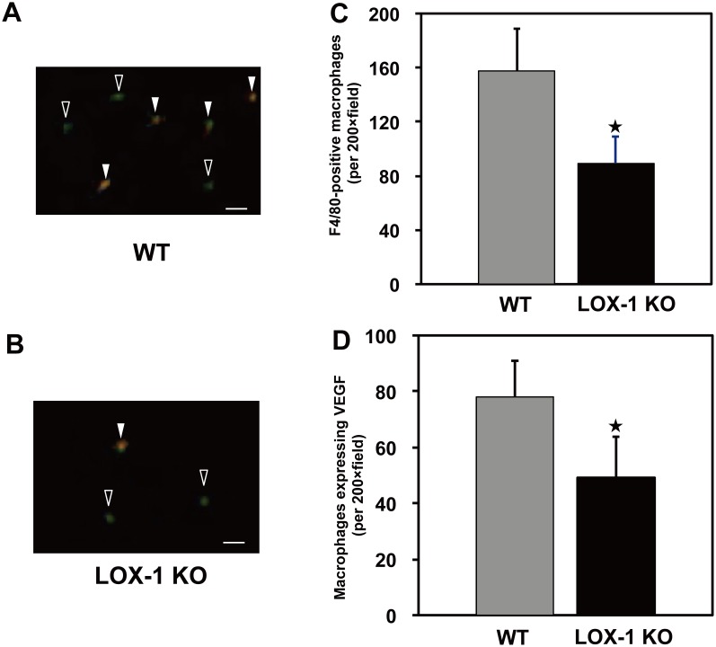
Figure 5. Two-color immunofluorescence staining.
Infiltrated macrophages (green) and VEGF protein (orange) in ischemic gastrocnemius muscle sections on postoperative day 3 after surgery. In the merged pictures (A and B), there were some overlaps between macrophage infiltration and VEGF expression, suggesting that these infiltrated macrophages produce and release VEGF protein. Quantitative data indicated both macrophage infiltration (C) and macrophages expressing VEGF (D) and both of these were significantly less in LOX-1 KO mice than in WT mice. Open arrow: macrophage without VEGF expression (green), Closed arrow: macrophage (green) with VEGF expression (orange), Original bars = 50 µm. ★, P<0.01 vs. WT, n = 20.

