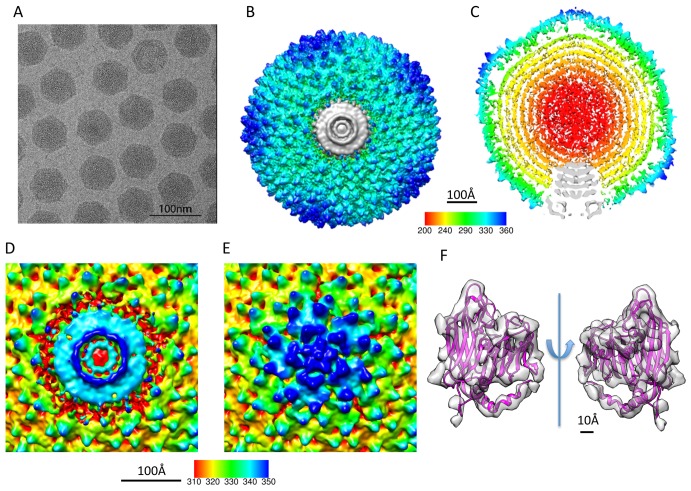
Figure 1. Reconstruction of PRD1 virion at 12 Å resolution without icosahedral symmetry imposition.
(A) A typical micrograph of the PRD1 virions. (B) The top view and (C) the central slice view of the reconstruction. (D) The unique vertex occupies one of the 12 pentonal positions and interacts with the capsid proteins at its outer edge. (E) The other 11 vertices have a regular 5-fold structure. (F) The crystal structure of the MCP P3 of PRD1 (PDB code 1W8X, chain B) fitted in the corresponding segmented density in the cryo-EM map, allowing the boundary delineation of the unique vertex complex and the MCPs surrounding it.

