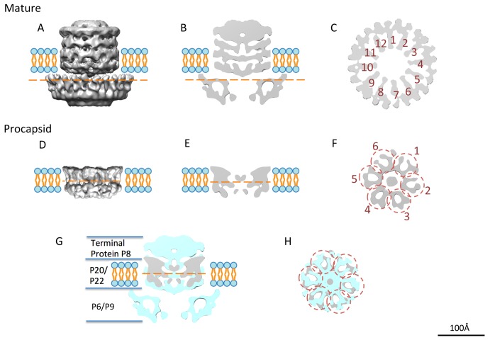
Figure 5. The unique vertex organization.
(A) The side view, (B) the central slice view, and (C) the top slice view of the segmented unique vertex in the mature virion. The cut-through view (C) (location labeled as the orange dashed line in (A) and (B)) shows the 12 arms (numbered 1 to 12) of the central part of the unique vertex and the extra surrounding densities. (D) The side view, (E) the side slice view, and (F) the top slice view of the segmented transmembrane density in the procapsid. The top slice view (F) (location labeled as the orange dashed line in (D)) shows the 12 arms of the structure organized as hexameric dimers (numbered 1 to 6 in circles). The density of the unique vertex in the procapsid (grey) is shown on that of the mature virion (cyan) as a side slice view (G) and top slice view (H).

