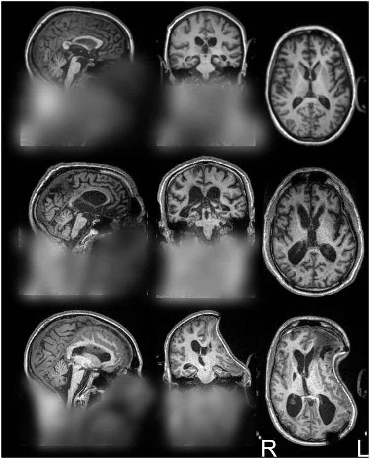
Figure 3. Sample patients’ full head images.
Orthogonal projection view of the original full head images for the three patients employed in Fig. 2. From top to bottom the images exemplify patient with relatively low, medium, and high brain pathology – relative to this cohort of patients. (Images are displayed in radiological convention; blurring added to protect the privacy of the patients).

