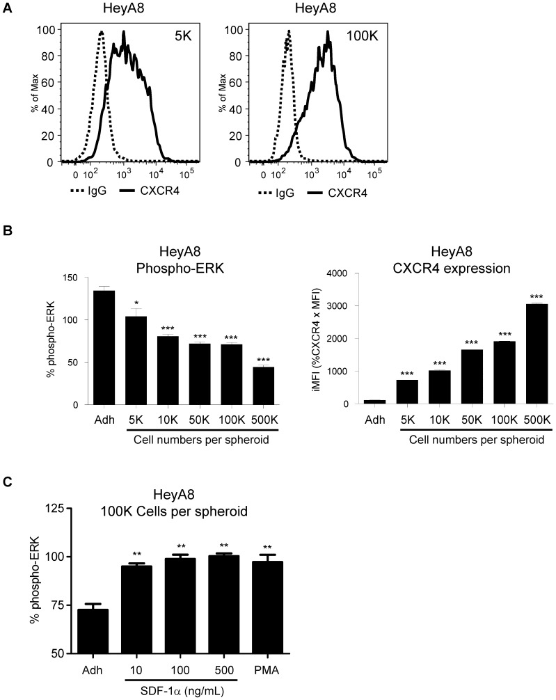
Figure 3. Phospho-ERK level is decreased in 3D spheroids.
HeyA8 cells were cultured as 3D spheroids with different number of cells per spheroid. (A) At 5000 cells per spheroid, there was a broad shift in CXCR4 surface expression. At higher cell numbers per spheroid, the expression levels of surface CXCR4 increased. (B) Corresponding normalized pERK levels showed a strong correlation between increasing CXCR4 surface expression with decreasing phospho-ERK levels. (*, P <0.05 vs Adh, 1-way ANOVA) (***, P <0.0001 vs Adh, 1-way ANOVA). (C) At 100,000 cells per spheroid, HeyA8 cells treated with SDF-1α and PMA showed activation of phospho-ERK levels. (**, P <0.005 vs Adh, 1-way ANOVA). Each bar graph is representative of 3 experiments.

