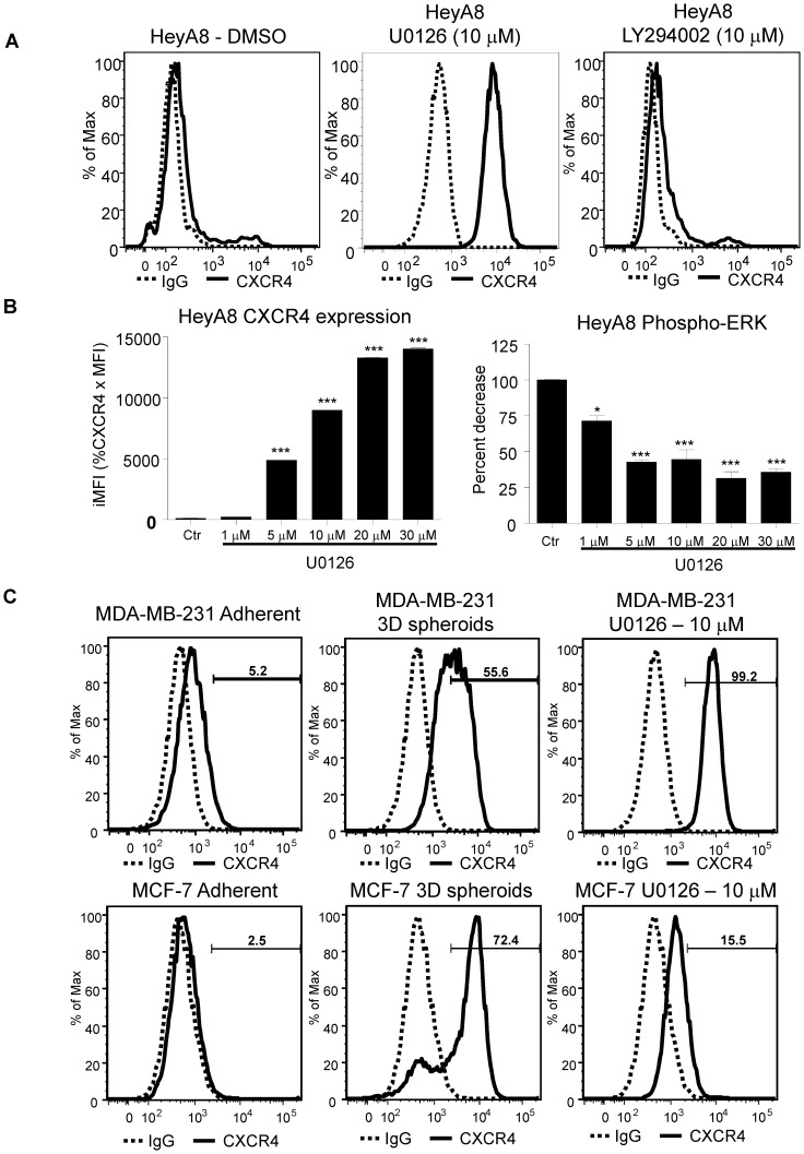
Figure 4. Phospho-ERK inhibition increases CXCR4 surface expression.
HeyA8 cells were cultured under normal adherent conditions. 100,000 HeyA8 cells were treated with MEK1/2 inhibitor U0126 and LY294002 at 10 µM for 48 hours. Cells were harvested and analyzed by flow cytometry. (A) CXCR4 expression was increased when treated with MEK1/2 inhibitor U0126 as compared to DMSO treated (solid lines). Similar to DMSO treatment, there was no detectable surface CXCR4 expression when treated with PI3K inhibitor LY294002. (*, P <0.05 vs Adh, 1-way ANOVA) (***, P<0.0001 vs Adh, 1-way ANOVA). (B) 200,000 HeyA8 cells were plated onto two 6 well plates and treated with U0126. One plate was harvested for CXCR4 expression and the other for phospho-ERK assay. The phospho-ERK assay showed decreasing levels with increasing U0126. Flow cytometry analysis showed a dose dependent increase in CXCR4 expression. (C) MDA-MB-231 and MCF-7 cells either grown as 3D spheroids or treated with U0126 showed increase CXCR4 expression similarly to HeyA8 cells. Results are representative of 3 experiments.

