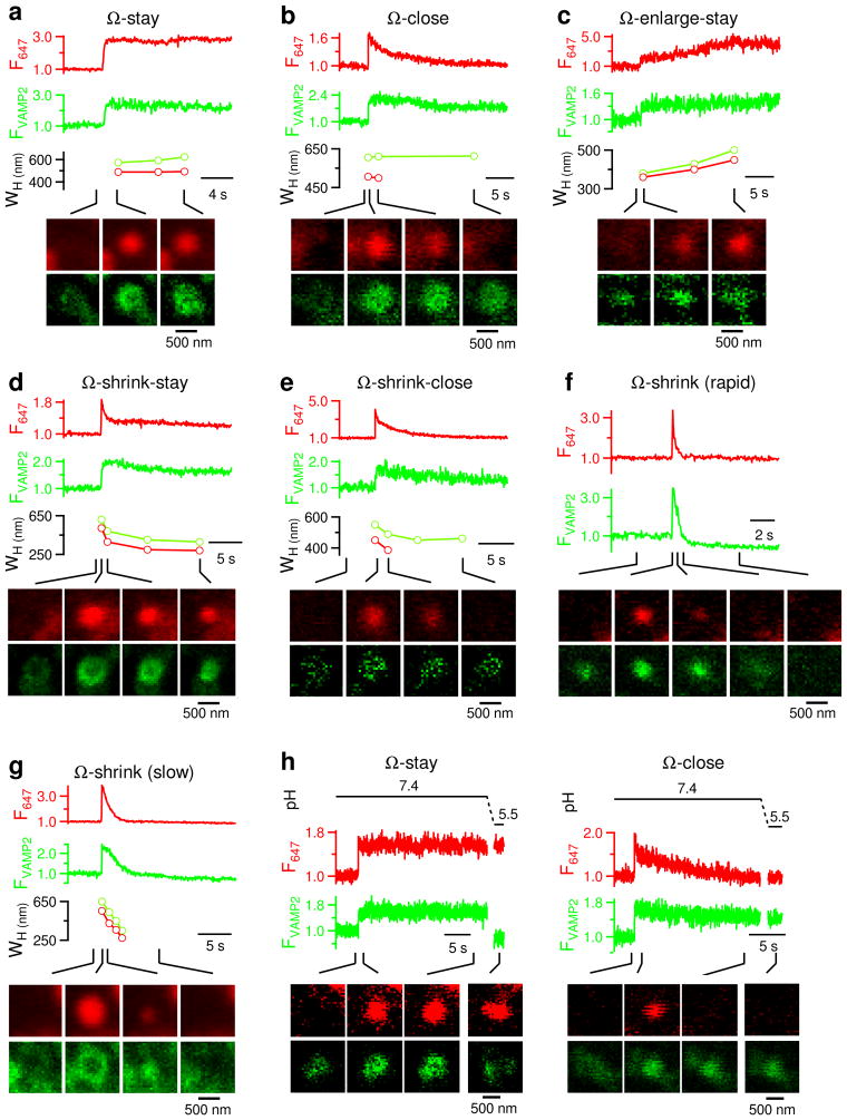
Figure 10. Ω-profile retains vesicle membrane protein VAMP2.
(a–g) F647 (red), FVAMP2 (green), WH of A647 (red) and VAMP2-EGFP (green) spot, and sampled A647 (red) and VAMP2-EGFP (green) images (at times indicated with lines) for spots undergoing Ω-stay (a), Ω-close (b), Ω-enlarge-stay (c), Ω-shrink-stay (d), Ω-shrink-close (e), and Ω-shrink (f: rapid shrinking, diffusion cloud; g: slow shrinking, size reduction observed). Cells were expressed with VAMP2-EGFP and stimulated by depol1s with A647 in the bath. WH is not measured in panel f, because VAMP2-EGFP rapidly diffused into a cloud, which did not reflect the Ω-shaped membrane profile size. VAMP2-EGFP spots appeared slightly (~50–100 nm in WH) larger than corresponding A647 spots (e.g., Fig. 10a–b), because VAMP2-EGFP was located at the membrane, whereas A647 was inside the Ω-shaped structure.
(h) The F647 and FVAMP2 changes in response to a bath pH change from 7.4 to 5.5 (upper) for spots undergoing Ω-stay (left) and Ω-close (right).

