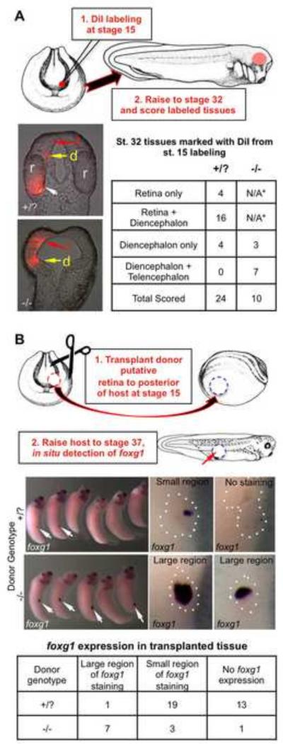
Figure 6. Presumptive retinal tissue is transformed into both diencephalic and telencephalic fates in rax mutant embryos.
(A) Retinal rudiment was labeled in St. 15 embryos using DiI marking. Embryos were then raised to St. 32 and scored for the fate of labeled tissues. Frontal sections of St. 32 embryos show DiI marking the retina (r, white arrow) and small amount of diencephalon (d, yellow arrow) in wildtype embryos, but not the telencephalon (t, red arrow). In mutant embryos, labeled tissues are found extensively in both the diencephalon and expanded telencephalon. Results shown in the table. *Since eyes fail to form in rax mutants, DiI labeling in the retina is not applicable (N/A). (B) Presumptive retinal tissue from St. 15 rax mutant or wildtype donor embryos was transplanted to the posterior of host embryos, which were raised to St. 37 and fixed. Donor embryos were allowed to develop in order to determine donor phenotype. In situ hybridization to detect telencephalic marker foxg1 was performed on hosts with donor tissue to determine the extent of telencephalic character in donor transplants. Left panels show lateral views of whole embryos, white arrows indicate regions positive for foxg1 expression; right panels show higher magnification view of transplanted region (outlined with white dots). Representative examples of each category (large or small region of transplant that is positive for foxg1expression domain relative to entire transplant size) are shown in right panels. Results from scoring are tallied in the bottom table.

