
Figure 1.
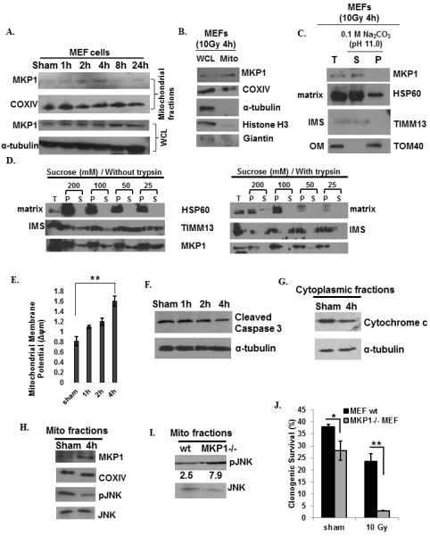
Mitochondrial MKP1 is enhanced by genotoxic stress in MEFs to inhibit mitochondria-initiated apoptosis via reduction of pJNK. A, Mitochondrial translocation of MKP1 in sham or 10 Gy irradiated MEFs. COXIV was used as the marker for mitochondria and IκB was used as the cytoplasmic marker. B, The purity of mitochondrial preparations in these experiments were further analyzed by immunoblots of MKP1, COXIV, α-tubulin (cytoplasmic marker), Histone H3 (nuclear marker), and Giantin (Golgi marker). C, Sub-mitochondrial localization of MKP1 detected by alkaline extraction (33). Total input (T), soluble matrix proteins (S), and membrane pellets (P) were immunoblotted for MKP1, TOM40 (an outer membrane protein), TIMM13 (an inter-space protein), and HSP60 (a matrix protein). D, Sub-mitochondrial localization of MKP1 detected via mitoplasting and protease protection assay (49). The total (T), pellet (P), and supernatant (S) fractions were subjected to western blotting with indicated antibodies. m (E, measured by fluorescent probe JC-1; n=3, **p<0.01), Caspase 3 cleavage (F), and cytochrome c release (G) in sham or 10 Gy irradiated MEFs. H, Increased MKP1 and decreased pJNK levels in mitochondrial fractions 4h post 10 Gy IR. I, JNK phosphorylation in the mitochondria of MKP1 knock-out and wt MEFs 4h after 10 Gy of radiation. pJNK levels were normalized to that of JNK levels and represented under the blots. J, Clonogenic survival analysis of wt versus MKP1−/− MEFs (n=3, *p<0.05, **p<0.01).