Figure 2.
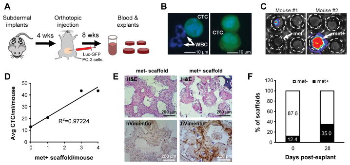
Implanted scaffolds capture circulating tumor cells and sustain metastatic engraftment ex vivo. A, schematic of experimental procedure. B, representative images of circulating tumor cells (CTCs; GFP+ cells) captured by microfluidic CTC-iChip. White blood cells (GFP−) are indicated by arrows. C, bioluminescence imaging (BLI) of explanted scaffolds confirmed CTC engraftment in some of the scaffolds. D, whole blood from 13 animals was pooled into three separate groups depending on the number of luciferase-positive scaffolds/mouse, either 0 (6 mice), 1 (5 mice) or >1 (2 mice) and run on CTC-iChips. The number of met+ scaffold/mouse was then plotted against the average number of CTC/ml/mouse showing linear correlation. E, histological analysis of met− and met+ scaffolds by human-specific Vimentin. F, after 4 weeks of ex vivo culture, 35% of scaffolds exhibited positive BLI signal (n=40).

