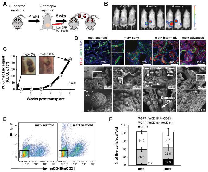
Figure 3.
Serial transplantation of scaffolds into naïve mice enables tracking of metastatic progression in scaffold microenvironments. A, scaffolds from primary orthotopic prostate tumor-bearing mice were transplanted into tumor-free syngeneic mice for long-term monitoring of metastatic progression. B, representative BLI images after serial transplant showing metastatic growth in the scaffolds. C, increased BLI signal from met+ scaffolds with gross images before and after serial transplant. Values are expressed as the R.L.U. (relative luciferase unit) sum of all positive scaffolds at any given time (number of met+ scaffolds indicated in the white circles; n=68). D, representative images of metastatic progression shown by immunofluorescence (top; blue=DAPI, green=mCD31, red=hVimentin) and SEM images of scaffold microenvironments (bottom). The scaffold pore composition includes hematopoietic, stromal and tumor cells with evidence of tumor/stromal interaction and local ECM degradation. Boxed areas in the middle panels are magnified in the lower panels. E, fluorescence activated cell sorting of cells recovered from met− and met+ PC-3 scaffolds. Boxed areas show stromal cells (GFP−/mCD45−/mCD31−). F, comparison of the relative cellular composition of met− vs met+ scaffolds from the PC-3 model by flow cytometry using the indicated antibodies. Bars represent SD (n=3).

