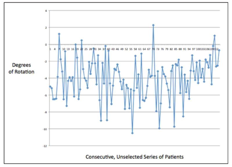
Figure 3.

Line graph demonstrating the variability in the relationship between the KAA and the TEA in a consecutive series of unselected patients undergoing TKA. KAA = posterior femoral axis of the kinematically aligned TKA, TEA = transepicondylar axis.
