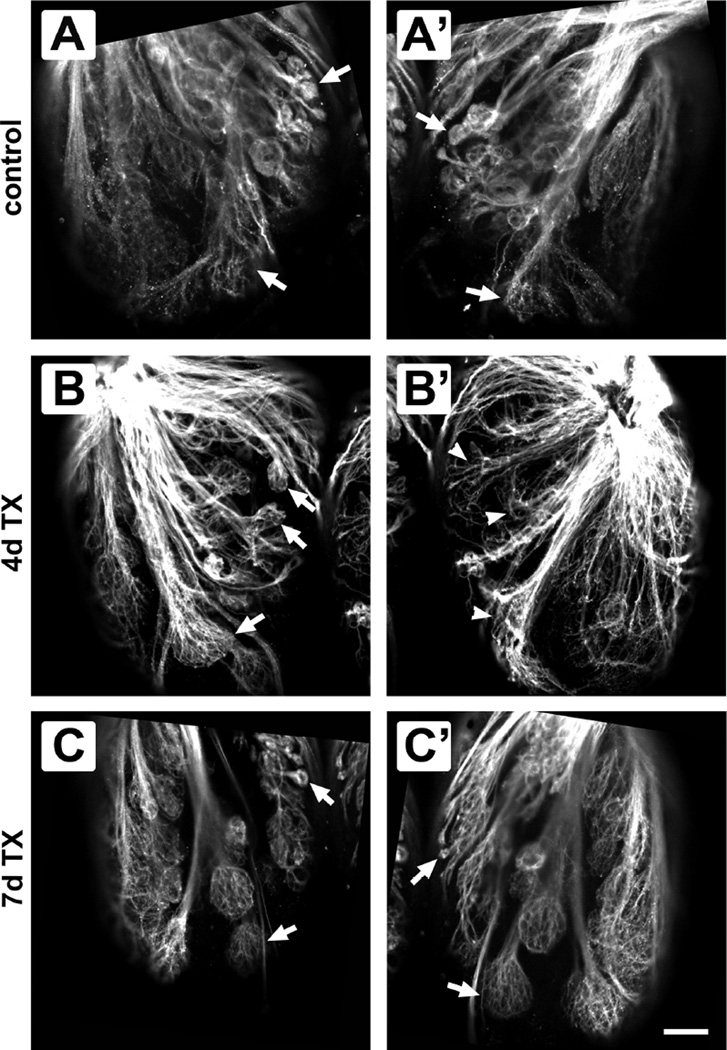
Figure 2.
Comparison of ventral glomerular patterns between olfactory bulbs of control and Triton X-100-treated fish. In untreated control fish, glomeruli (arrows) appeared the same between the left (A) and right (A’) olfactory bulbs. Four days after detergent ablation of the olfactory epithelium, glomeruli in the treated bulb (B’) seemed disrupted when compared to the internal control bulb (B). Even with this disruption, the same glomeruli in the untreated bulb (arrows in B) could be identified in the experimental bulb (arrowheads in B’). Seven days after treatment, glomeruli in the internal control side (C) and the treated side (C’) had similar morphology, and the same glomeruli were seen in both olfactory bulbs (arrows). Scale bar = 50 µm for all.

