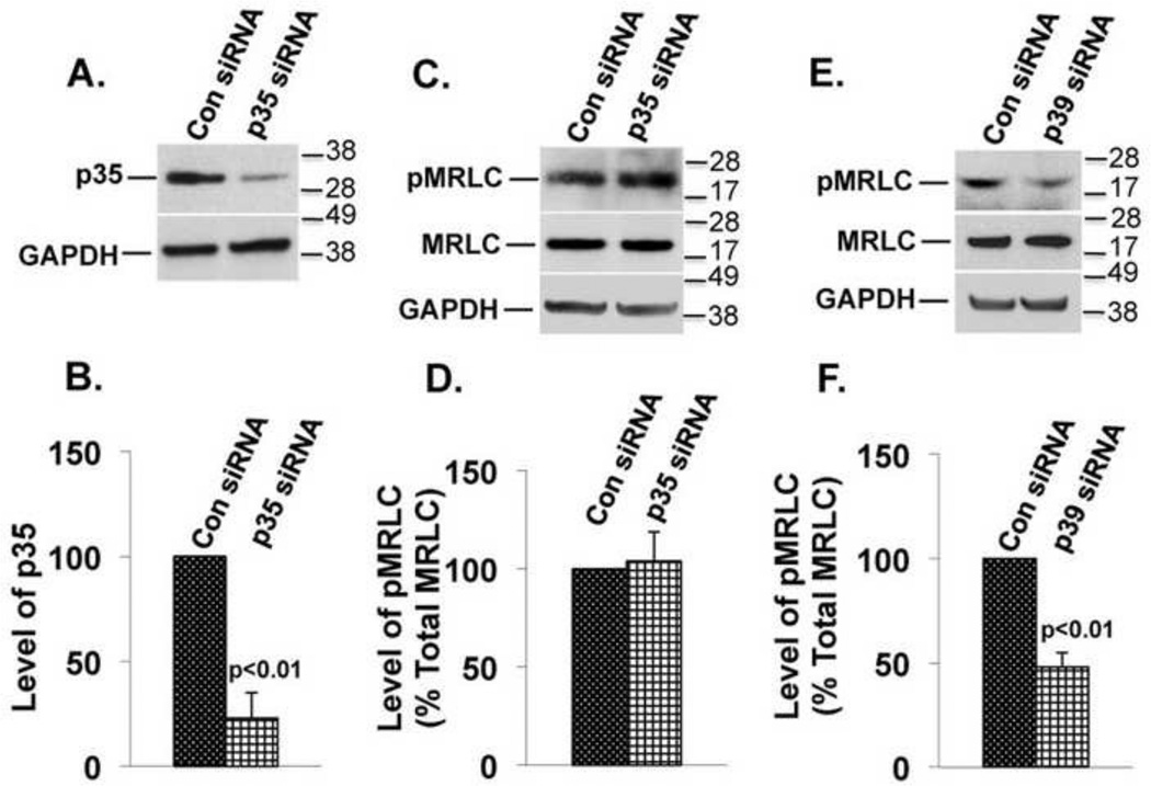
FIGURE 7. p39 siRNA but not p35 siRNA reduces MRLC phosphorylation.
(A) Suppression of p35 protein expression by p35 siRNAs (upper panel) and GAPDH was used as a loading control (lower panel). (B) The results of three independent experiments as in A were quantified by densitometry and normalized to GAPDH. The graph represents mean ± s.e.m. (C) The experimental conditions were similar to those in A. Immunoblots for pMRLC (upper panel), total MRLC (middle panel), and GAPDH (lower panel). (D) Results of three independent experiments as shown in C were quantified by densitometry, and the ratio of pMRLC to total MRLC was plotted. The values represent mean ± s.e.m. Suppression of p35 did not alter the level of pMRLC. (E) The experimental conditions were similar to those in A except p39 siRNAs was transfected. Immunoblots for pMRLC (upper panel), total MRLC (middle panel), and GAPDH (lower panel) are shown. (F) Results of three independent experiments as shown in E were quantified by densitometry, and the ratio of pMRLC to total MRLC was plotted. The values represent mean ± s.e.m. The level of pMRLC was reduced (p<0.01) in cells transfected with p39 siRNAs as compared to cells transfected with control siRNA.

