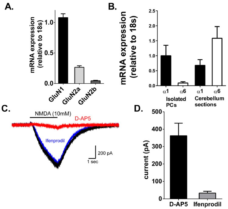
Figure 6.

NMDA receptors in Purkinje cells are predominantly GluN1/GluN2A containing receptors. (A.) Relative expression of GluN1 subunits in Purkinje cells. mRNA expression in Purkinje cells isolated by laser capture microdissection were quantified relative to the housekeeping gene 18s and normalized to GluN1 expression. Bars represent mean±SEM (n=4) (B) GABAA α6 mRNA was expressed at very low levels in LCM samples, demonstrating an enrichment of Purkinje cells with few granule cells being collected. GABAA α1 mRNA was observed in both LCM and whole cerebellar sections. N=4. (C.) Representative traces of antagonist-sensitive current elicited by iontophoresis of NMDA (10 mM, 4s). Currents elicited in the presence of antagonist were subtracted from baseline currents to generate antagonist-sensitive current. Solid line indicates timing of NMDA iontophoresis. (D.) Summarized data of current (in pA) blocked by D-AP5 (n=7) and ifenprodil (n=5). Bars represent mean±SEM.
