
Figure 4. Therapeutic efficacy of a pH-sensitive TAT-Au NP-Dox delivery system in an intracranial U87 glioma mouse model.
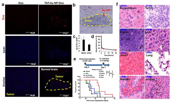
(a) Confocal images of mouse brain tissues 24 hours after I.V. injection showed Dox accumulation in a mouse after TAT-Au-NP administration. No Dox accumulation was observed when the drug alone was administered (b) Brain tissue from a TAT-Au NP-Dox injected mouse stained by H&E and silver enhancement confirming the localization of TAT-Au NP on the brain tumor tissue. (c) Quantification of the gold contents in the brain and urine samples from TAT-Au NP-Dox treated mice (n=5). (d) Quantification of the gold in the blood samples from TAT-Au NP-Dox injected mice. (e) Kaplan-Meier survival curves of intracranial U87 glioma tumor bearing mice I.V. injected with normal saline (n=5), Dox (n=6), Au NP-Dox (n=4) and (f) TAT-Au NP-Dox (n=6) at Dox concentration of 1.5 mg/kg. (f) Histology images of tissue samples with H&E and silver enhancement staining from the TAT-Au NP-Dox-treated mouse post 6 weeks of injection.