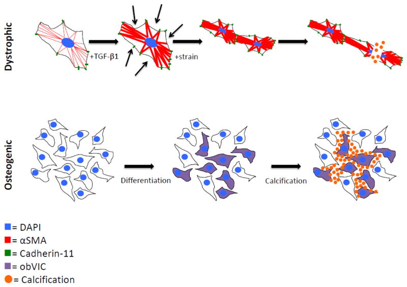
Figure 1.
Cartoons depicting proposed mechanisms of valve calcification. The dystrophic pathway is mediated by a TGF-β1 mediated increase in αSMA and cadherin-11, which increases the cells’ contractility and strengthens their connections to each other. Under pathological strain, the increased and uneven tension tears cells apart, leading to calcification via apoptosis. The osteogenic pathway proceeds by osteogenic differentiation into obVICs, likely from qVICs. These obVICs actively form mineralized deposits.

