
Figure 1.
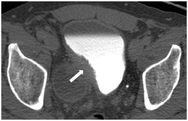
Male age 53 years. CT urography scan shows a large soft tissue mass involving the diverticulum neck and right lateral wall of the bladder. The mass extends into the extravesical fat (arrows). The patient was found to have transitional cell carcinoma with sarcomatoid features, stage T3.