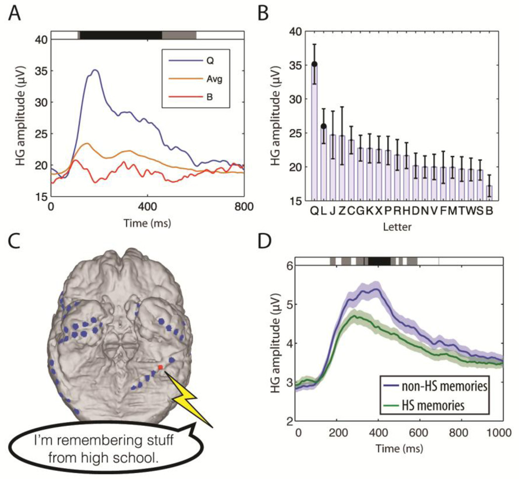
Fig. 7. Electrocorticography reveals the neural basis of memories.
(A) The amplitude of high gamma (HG; 65–150 Hz) ECoG activity at a site in one patient’s ventral temporal lobe in a working memory task (Modified from Jacobs and Kahana 2009). Shading at top indicates time points that exhibited significant differences in amplitude according to the identity of the viewed item. (B) The amplitude of HG activity for all letters viewed in the task (same electrode as panel A.) (C) Brain image from a patient who spontaneously remembered memories of high school (HS) after stimulation at the indicated electrode (red) in his left ventral temporal lobe. (D) The amplitude of HG activity observed from this electrode when the patient performed a memory task where he remembered HS and non-HS information (same electrode as panel C.)

