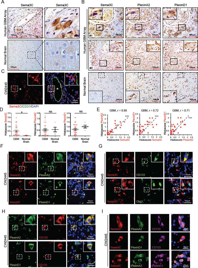
Figure 1. Sema3C and Its Receptors Are Co-expressed In Stem Cell Marker+ GBM Cells.
(A and B) Immunohistochemical (IHC) staining of Sema3C, PlexinA2 and PlexinD1 in serial sections of human GBM tissue array. Sections were counterstained with hematoxylin. Asterisk denotes vessel lumen.
(C) Immunofluorescent (IF) staining of Sema3C (red) in relation to blood vessels marked by CD31 staining for endothelial cells (green) in human primary GBM tissues. * denotes vessel lumen.
(D and E) Histoscores (D) and correlation analysis (E) of human GBM tissue array stained for Sema3C, PlexinA2 and PlexinD1. *p< 0.05, ***p < 0.001.
(F) IF staining of Sema3C and PlexinA2/D1 on frozen sections of human primary GBM. Nuclei were counterstained with DAPI (blue).
(G–I) IF staining of Sema3C, PlexinA2/D1 and GSC markers CD133 and Olig2 on frozen sections of human primary GBM. Nuclei were counterstained with DAPI (blue).
See also Figure S1.

