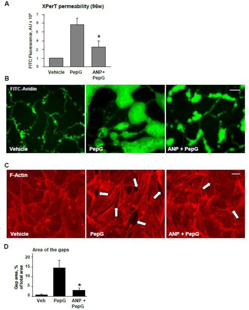
Figure 1. ANP attenuates PepG-induced EC permeability and actin remodeling.
HPAEC grown in 96-well plates (A) or on glass coverslips (B, bar=10 µm) with immobilized biotinylated gelatin (0.25 mg/ml) were pretreated with ANP (100 nM, 20 min) followed by challenge with PepG (0.25 µg/ml, 6 hrs), and addition of FITC-avidin (25 µg/ml, 3 min). Unbound FITC-avidin was removed, and FITC fluorescence was measured; normalized data are expressed as mean ± SD; *P <0.05 vs. PepG alone. C: EC grown on glass coverslips were pretreated with ANP (100 nM, 20 min) followed by challenge with PepG (0.25 µg/ml, 4 hrs) and immunofluorescence staining with Texas Red phalloidin to detect actin filaments. Intercellular gaps are shown by arrows; bar=10 µm. D: Bar graphs depict quantitative analysis of total gap area in control, PepG and ANP + PepG treated EC monolayers; data are expressed as mean ± SD; *P <0.05 vs. PepG alone.

