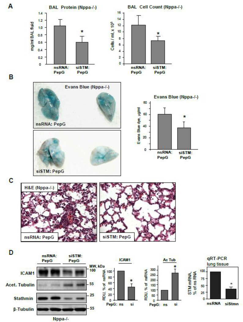
Figure 10. Stathmin knockdown attenuates the exacerbation of PepG-induced lung injury in ANP-deficient mice.
Control and ANP knockout mice were transfected with non-specific or stathmin-specific siRNA for 72 hrs followed by treatment with PepG for 24 hrs. A: Protein concentration and total cell count were performed in BAL samples. B: Lung vascular permeability was assessed by Evans blue accumulation in the lung tissue; n=4 per condition; data are expressed as mean ± SD; *p<0.05 vs. nsRNA. B: Morphological changes in lungs from PepG-challenged control and ANP-deficient mice were evaluated by hematoxilin and eosin staining of lung tissue sections. D: ICAM-1 expression and acetylated tubulin levels in lung tissue lysates were measured by western blot (left panel). Western blot detection of β-tubulin in corresponding tissue lysates was used as a normalization control. Bar graphs depict quantitative densitometry analysis of western blot data; data are expressed as mean ± SD; *P <0.05 vs. nsRNA. Stathmin knockdown in lung tissue was confirmed by qRT-PCR (right panel).

