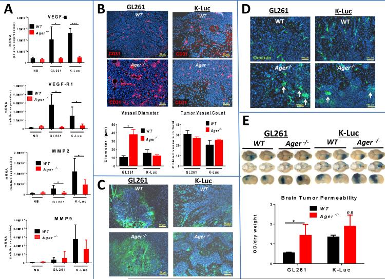
Figure 4.
RAGE ablation abrogates tumor angiogenesis. A, qPCR of normal brain (NB) and intracranial GL261 and K-Luc gliomas demonstrating lower expression of tumor proangiogenic factors in Ager−/− mice (n=3 mice/group ± SD). B, Morphology (top panel) and quantification (lower panel) of GL261 and K-Luc blood vessels in WT and Ager−/− mice (n=4 mice/group ± SD). C, Tumor hypoxia (green staining) was measured by injecting glioma-bearing mice with pimonidazole two hours prior to tissue examination. Intracranial tumors were harvested two weeks after implantation. D, Effect of RAGE ablation on vascular permeability. Mice bearing two-week old intracranial GL261 or KLuc were injected with TRITC-dextran 150 two hours prior to image analysis. Vascular leakage is evident as dextran extravasation into perivascular space (arrows). E, Tumor vascular permeability was also assessed with Evans blue assay. Tumor-bearing mice were injected with Evans blue dye 30 minutes prior to sample collection. The dye was extracted and quantified with a spectrophotometer. (n=4 mice/group ± SD). *: p<0.05, ***: p<0.001. Experimental results are representative of two separate experiments.

