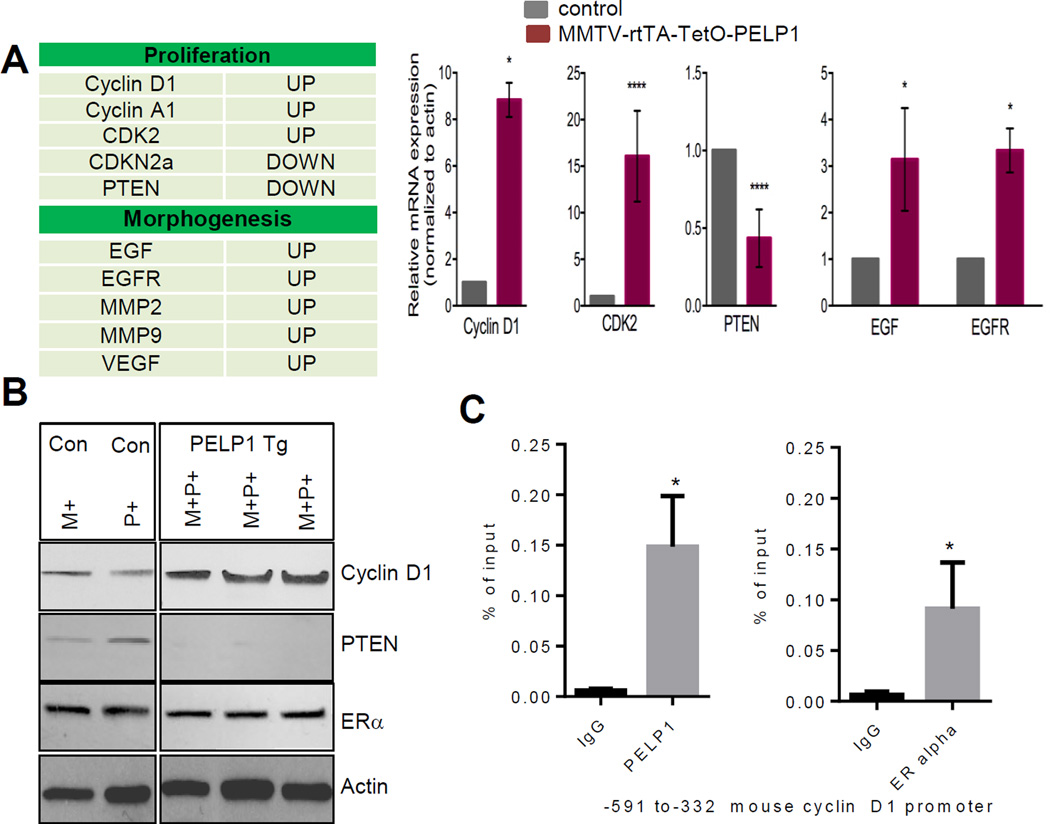
Figure 5.
Effect of PELP1 transgene expression on the expression of breast cancer pathway genes in the mammary gland. A, Gene profile of PELP1-mediated alterations in genes in the mammary gland was analyzed by using real-time RT2 ProfilerTM pathway–focused PCR array. Total RNA isolated from 6-month-old control and MMTV-rtTA-TetO-PELP1 mice was used. The list of the top five genes is shown in the table. Validation of five representative genes was done by using real-time RT-PCR. All experimental data points were generated from three biological replicates per transgenic line (left panel). Statistical significance was determined by using a Student’s t test.****p<0.0001; *p<0.05. B, Western blot analyses of the status of cyclin D1, PTEN and ER-alpha in control and MMTV-rtTA-TetO-PELP1 mammary glands (left panel). Actin was used as control. WT, wild type; M+ MMTV-rtTA transgenic mice; P+ TetO-PELP1 mice ; and M+P+, MMTV-rtTA-TetO-PELP1 bitransgenic mice. C, ChIP assay was done using the DNA isolated from the mammary glands of 6 months old doxycycline-treated PELP1 bitransgenic animals with antibodies specific for PELP1, ER or the isotype IgG control. DNA recovered from ChIP or input controls was subjected to real-time quantitative PCR with primers specific to −591 to −332 that represent the proximal promoter regions of mouse cyclin D1 promoter. Statistical significance was determined by using a Student’s t test. *p<0.05.

